The spinal canal is the anatomic casing for
the spinal cord. The spinal cord extends in the adult from the top of the
Atlas (first vertebrae) to the lower border of the first lumbar vertebrae
(L1). There it divides into many different nerve roots, which are called Cauda
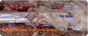
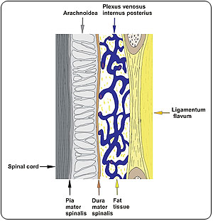
equina. The spinal cord is enclosed in a rather stiff dural sheath, called
Dura mater spinalis (thickness ~2mm). The space between dura and vertebral
body is called epidural space. It is filled with ligaments (Ligamentum flavum,
Ligamentum longitudinale posterius), fat
tissue
and a plexus venosus internus, which provides the venous drainage for the
individual vertebrae of the spinal column. Consequently, PADeMIS has only
a limited space (4-15 mm diameter) inside the epidural space. PADeMIS has
to produce as much as 2N in order to generate a forward motion.
For morphometry, the different components of the epidural space have to be
measured with defined accuracy. Presently, preparations, Magnetic Resonance
Imaging (MRI) and Computed X-ray Tomography (CT) scans of humans, dogs and
pigsare analysed for this purpose.